THE ENDOCRINE SYSTEM
The endocrine system is a complex network of chemical signals and messages that control many immediate and life-long bodily responses and functions. Growing taller, developing male or female characteristics and reacting to fear are all partially directed by endocrine hormones. All animals with backbones - from fish to mammals - have an endocrine system that works hand-in-hand with the nervous system to:
maintain the body's internal steady state (nutrition, metabolism, excretion, water and salt balance);
react to stimuli from outside the body;
regulate growth, development and reproduction; and
produce, use and store energy.
The endocrine system's three parts - glands, hormones and target cells - relay information and instructions throughout the body. Sometimes the whole process works within seconds, say, in response to fear. Other times it reacts more slowly, telling body parts when and how much to grow and developing characteristics that distinguish male from female.
The endocrine system is made up of specialized cells, glands and hormones. Acting like a communication network, it responds to stimuli by releasing hormones, the chemical messengers that carry instructions to target cells throughout the body, from endocrine glands. The target cells read and follow the hormones' instructions, sometimes building a protein or releasing another hormone. These actions lead to many bodily responses such as a faster heart beat or bone growth.
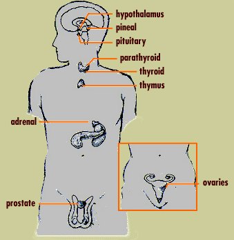
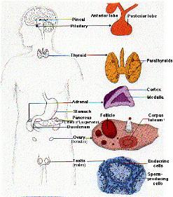
All vertebrate animals (fish, amphibians, reptiles, birds and mammals, including humans) have the same endocrine glands and release similar hormones to control development, growth, reproduction and other responses. Here are some of the main glands.
- Hypothalamus
- Pineal gland
- Anterior pituitary gland
- Posterior pituitary gland
- Thyroid
- Parathyroid gland
- Thymus
- Adrenal glands (medulla and cortex)
- Pancreas
- Ovary (follicle and corpus luteum)
- Testis
Melatonin and the Pineal Gland
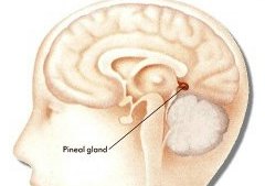
The pineal gland is a tiny structure located at the base of the brain.
Its principal hormone is melatonin, a derivative of the amino acid tryptophan.
Synthesis and release of melatonin is
stimulated by darkness and
inhibited by light.
But even without visual cues, the level of melatonin in the blood rises and falls on a daily (circadian) cycle with peak levels occurring in the wee hours of the morning.
The pineal gland is a tiny organ in the brain of human beings and most other vertebrates (animals with a backbone). Scientists are uncertain of the function of the pineal gland in human beings. They believe it plays a role in a variety of important body functions, including certain reproductive processes. In most other vertebrates, the pineal gland helps regulate certain daily and seasonal body cycles by secreting a hormone called melatonin.
Production of melatonin varies with periods of light and darkness in the environment. Even though in human beings the pineal gland lies near the centre of the brain, it obtains information about light in the environment through nerve pathways originating in the eyes. In general, light slows and darkness stimulates the pineal gland's production of melatonin. Therefore, the gland tends to secrete small amounts of melatonin during the day and larger amounts at night.
In human beings, melatonin has been linked to the onset of puberty, the stage of life when a person matures sexually. Studies have shown that the pineal gland's nightly secretion of melatonin decreases when a boy or girl reaches puberty. Other studies have indicated that melatonin may help regulate menstrual cycles in women and sperm production in men. In addition, researchers have suggested a connection between melatonin levels and certain mental illnesses.
It would be difficult to overstate the influence of hypothalamic and pituitary hormones over physiologic processes. The target cells for most of the hormones produced in these tissues are themselves endocrine cells, and a seemingly small initial signal is thus amplified to cause widespread effects on many cells and tissues.
The close anatomical and functional relationships between hypothalamus and pituitary force an integrated discussion of these organs. The focus here is to introduce the major hormones produced by these organs, with significant emphasis on how hormone secretion and action are controlled. Links are provided to other sections of text containing additional information on the effects of these hormones.
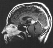
The hypothalamus is a region of the brain that controls an immense number of bodily functions. It is located in the middle of the base of the brain, and encapsulates the ventral portion of the third ventricle.
This pattern of vascular connections is presented diagrammatically below.
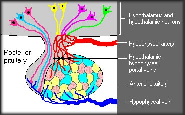
The utility of this unconventional vascular system is that minute quantities of hypothalamic hormones are carried in a concentrated form directly to their target cells in the anterior pituitary, and are not diluted out in the systemic circulation.
The pituitary gland is often portrayed as the "master gland" of the body. Such praise is justified in the sense that the anterior and posterior pituitary secrete a battery of hormones that collectively influence all cells and affect virtually all physiologic processes.
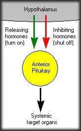
The pituitary gland may be king, but the power behind the throne is clearly the hypothalamus. As alluded to in the last section, some of the neurons within the hypothalamus - neurosecretory neurons - secrete hormones that strictly control secretion of hormones from the anterior pituitary. The hypothalamic hormones are referred to as releasing hormones and inhibiting hormones, reflecting their influence on anterior pituitary hormones.
Hypothalamic releasing and inhibiting hormones are carried directly to the anterior pituitary gland via hypothalamic-hypophyseal portal veins. Specific hypothalamic hormones bind to receptors on specific anterior pituitary cells, modulating the release of the hormone they produce.
As an example, thyroid-releasing hormone from the hypothalamus binds to receptors on anterior pituitary cells called thyrotrophs, stimulating them to secrete thyroid-stimulating hormone or TSH. The anterior pituitary hormones enter the systemic circulation and bind to their receptors on other target organs. In the case of TSH, the target organ is the thyroid gland.
Thyroid hormones affect three fundamental physiologic processes: cellular differentiation, growth, and metabolism. If you think about that statement for a minute, you might legitimately ask "So what else is there?" which emphasizes just how much of physiology is affected by thyroid hormones. Not many hormones can claim as diverse a set of target cells.
The thyroid gland also produces another hormone called calcitonin, and the parathyroid glands secrete parathyroid hormone. Parathyroid hormone and calcitonin participate in control of calcium and phosphorus homeostasis and have significant effects on bone physiology.
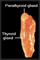
The thyroid gland is located in the neck, in close approximation to the first part of the trachea. In humans, the thyroid gland has a "butterfly" shape, with two lateral lobes that are connected by a narrow section called the isthmus. Most animals, however, have two separate glands on either side of the trachea. Thyroid glands are brownish-red incolor.
Close examination of a thyroid gland will reveal one or more small, light-colored nodules on or protruding from its surface - these are parathyroid glands (meaning "beside the thyroid"). The image above shows a canine thyroid gland and one attached parathyroid gland.
The microscopic structure of the thyroid is quite distinctive. Thyroid epithelial cells - the cells responsible for synthesis of thyroid hormones - are arranged in spheres called thyroid follicles. Follicles are filled with colloid, a proteinaceous depot of thyroid hormone precursor (more about that later).
In addition to thyroid epithelial cells, the thyroid gland houses one other important endocrine cell. Nestled in spaces between thyroid follicles are parafollicular or C cells, which secrete the hormone calcitonin.
The structure of a parathyroid gland is distinctly different from a thyroid gland. The cells that synthesize and secrete parathyroid hormone are arranged in rather dense cords or nests around abundant capillaries.
Back to Homepage
Edication - HowComYouCom.com
|